
Micro Anatomy of Eye
Product Code : JA-AM-6047
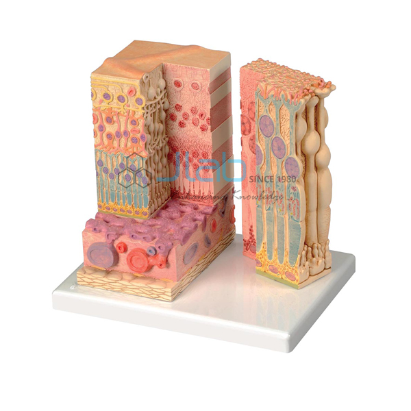
The Micro Anatomy of Eye Model from Jaincolab illustrates the microscopic structure of the retina with choroid and sclera.
This eye histology model has a block-like, layered left side showing the complete structure of the retina, including the supplying vascular layer and parts of the sclera, from a light-microscopic view. The right part of the model is a sectional enlargement showing the microscopic structure of the photoreceptor and the cells of the pigmented layer.
As a reliable biology lab equipment supplier in India, Jaincolab supplies this model to schools, colleges and biology laboratories across India and internationally.
Product Specifications
| Parameter | Details |
|---|---|
| Structures Shown | Retina, choroid, sclera |
| Left Side | Complete layered retina structure with vascular layer and sclera, light-microscopic view |
| Right Side | Sectional enlargement of photoreceptor and pigmented layer cells |
| Product Code | JA-AM-6047 |
Key Features
- Illustrates microscopic structure of the retina with choroid and sclera
- Left side shows complete layered retina structure
- Shows the supplying vascular layer and parts of the sclera
- Right side is a sectional enlargement
- Shows photoreceptor and pigmented layer cell structure
Why Choose Jaincolab
Jaincolab is a trusted biology lab equipment supplier in India, serving schools, colleges and biology laboratories across India and internationally. This model complements Jaincolab's gross-anatomy eye models within the wider Anatomical Models Equipments range. Jaincolab is ISO 9001:2015 and ISO 14001:2015 certified.
Applications
| Application Area | Use |
|---|---|
| Ophthalmic Histology Teaching | Study of retina, choroid and sclera microstructure |
| School & College Science Labs | Demonstration of photoreceptor and pigmented layer cells |
| Biology Classrooms | Reference model for eye tissue histology |
| Practical Examinations | Comparative study of retinal layers and vascular supply |
Frequently Asked Questions
Q1. What does this model illustrate?
A. The microscopic structure of the retina with choroid and sclera.
Q2. What does the left side of the model show?
A. The complete layered retina structure, including the vascular layer and parts of the sclera, from a light-microscopic view.
Q3. What does the right side of the model show?
A. A sectional enlargement showing the photoreceptor and pigmented layer cells.
Q4. Who is this model suitable for?
A. Students studying eye histology or ophthalmology at school, college or laboratory level.
Q5. Is this model related to Jaincolab's other eye models?
A. Yes, it complements Jaincolab's gross-anatomy eye models within the same Anatomical Models Equipments range.
Request a Quote
To request pricing or place an order for the Micro Anatomy of Eye Model, please contact our team with your requirement.


